The functional study of the brain requires the development of tools to record neuronal activity. The electrical currents that arise in neurons are chemical in nature, which makes techniques such as functional magnetic resonance imaging possible. In addition, the currents themselves generate weak electric and magnetic fields that, with appropriate equipment, can be recorded non-invasively, outside the head volume. Our focus is magnetoencephalography: a method that allows us to analyze neuronal electrical activity by recording the magnetic fields it generates.
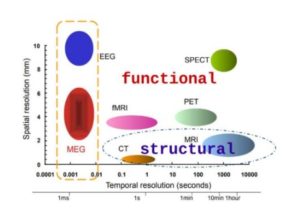
As a direct method of recording electrical activity, MEG provides high temporal resolution (about 1 ms). In addition, the transparency of biological tissues to magnetic fields allows obtaining high spatial resolution, supported by high sensitivity to high frequencies. Combining high temporal and spatial resolution with sensitivity to high-frequency signals, MEG is by right the most powerful non-invasive neuroimaging method.
Despite all the advantages, MEG has been and remains an unpopular method. This is due to the fact that traditionally MEG uses superconductor quantum interferometers (SCVID) as highly sensitive magnetometers. The operation of the latter is based on the phenomenon of low-temperature superconductivity, which imposes a number of limitations on the use of MEG due to the need to maintain sensors in the state of superconductivity in a thick-walled Dewar vessel with liquid helium. The disadvantage of such expensive and non-mobile systems is the great distance of the sensors from the brain, which significantly reduces the signal-to-noise ratio of the measured data. In addition, such systems require the use of an extremely expensive consumable, liquid helium, which is used to cool the sensors and keep the Josephsonian contacts operational.
In the last decade, however, new types of highly sensitive sensors applicable to MEG are emerging. These are optical pumped magnetometers (MON or OPM) and solid-state magnetometers based on iron-yttrium garnet films (YIGM or YIGM). These sensors operate at room temperature, are compact, and do not require liquid helium cooling, which removes much of the limitations of MEG as a method and could potentially make MEG a more reliable and less expensive instrument while retaining all of its advantages. The work of our group is largely devoted to the development of MEG systems based on these types of sensors.
The activity of the group is multidisciplinary and is aimed at the development of new neuroimaging techniques both in terms of mathematical and algorithmic support and hardware. In particular, one of the main directions of the group’s work is the development of new magnetoencephalography (MEG) systems using innovative highly sensitive magnetic sensors: optically pumped atomic magnetometers (OPAM) and solid-state magnetometers based on iron-yttrium garnet films (ISGM).
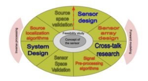
Building new magnetoencephalography systems involves working in the following directions:
It is worth noting that these directions are closely related to each other, and the results obtained in the work in one direction can and will be used in the work on other directions.
The work on creation of new types of MEG devices begins with work on the root element of any MEG system: a magnetic field sensor. So far, we have presented two types of such sensors: MON and LIGM. The applicability of each of these sensors in the context of MEG has been shown by us earlier (see [Petrenko et. al, 2021], [Koshev et. al, 2021]), which is the zero step, a precursor to the development of a MEG system. The next step is the development of no longer a sensor, but a sensor system, which can also lead to a new stage of refinement of the sensors themselves. The development of the sensor system is performed using methods of mathematical physics – signal modeling of sensor systems taking into account the properties of sensors, and includes both basic research in the field of efficiency of sensor systems and task-oriented optimization of positions and orientations of sensitive axes of sensors. An example of basic efficiency research is the development of sensor system efficiency metrics to determine the main direction for further sensor development: increasing sensitivity, minimizing intrinsic noise, or reducing size in order to arrange more sensors in a limited volume to capture a more detailed signal. The second study, task-oriented optimization of sensor systems, is based on the developed metrics for evaluating the efficiency of the system based on information theory and statistical methods of data processing (such as signal-to-noise ratio or sensitivity to sources in a particular brain region, Cramer-Rao estimates for the variance of the position of a localized source, etc.).

The development of an experimental paradigm for working with new types of sensors includes both research into the applicability of sensors in different environments, research into the infrastructure required for sensor operation, such as the creation of coils to actively suppress background magnetic fields and/or magnetic shields, and the relative positioning of sensors to minimize interference and magnetic interference. In addition, the sensors, of course, require some calibration or graduation, also part of the experimental paradigm development tasks.
Each of the sensors under development has its own characteristics leading to some, obvious or hidden, differences in the signal they record. The obvious differences include, for example, the differences determined by the geometry of the sensors and the orientation of their sensitive axes relative to the head, as well as the boards for collecting and recording the recorded signal. An example of less obvious differences is, for example, the inapplicability of standard algorithms for filtering the signal from external noise (e.g., Maxwell filter, [Taulu et. al, 2005]) to systems with a small number of sensors, which forces either to improve the properties of magnetic screens and active noise reduction systems, or to develop new algorithms for data preprocessing and filtering.
Inverse problem solving methods are used to form a map of activity on the cerebral cortex based on sensor signals. Another extremely important direction of the group’s work is research in the field of algorithms for solving the inverse problem of MEG – localization of electrical sources in the cerebral cortex, as well as approaches to the detection of functional networks.
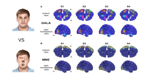
Both of these problems lack a unique mathematical solution and require the use of additional a priori information to achieve accurate and reliable results. The Figure shows an example of how the new GALA algorithm works in solving the inverse problem using an a priori assumption of weak similarity of brain activity between subjects.
Additionally, in the context of analyzing the activity of cortical sources, it is important to assess the functional connectivity between them, as determined by the constancy of phase delays in the synchronous activity of neuronal populations.
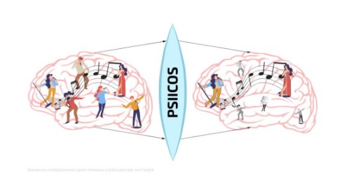
One of the innovative methods in this area is the PSIICOS (Phase shift invariant imaging of coherent sources) method developed by the Center for Bioelectrical Interfaces of the National Research University Higher School of Economics, with which LIFT closely cooperates within the framework of the MEG project. This method is specifically designed to assess functional connectivity independent of phase delays between the rhythmic activity of neuronal populations. The PSIICOS method can suppress volume conduction artifacts and for the first time has provided access to mapping functional networks with in-phase activity that are regularly reported in invasive measurements but have remained inaccessible to non-invasive neural mapping methods. In the musical allegory presented in the Figure above PSIICOS projection allows us to suppress the activity of single singers and emphasize the relative loudness of a melody sung by multiple singers synchronously.
LIFT together with the Center for Bioelectrical Interfaces at the Higher School of Economics is developing an alternative MEG system based on OPM sensors operating on the principle of optical pumping. Such sensors have a compact body and can be mounted on conventional EEG caps, providing freedom of movement and more accurate source localization compared to widely used SQUID systems.
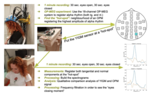
The project has successfully demonstrated the recording of functional brain rhythms such as occipital alpha rhythm and sensorimotor rhythm also in the ideomotor neurointerface paradigm. Optically pumped sensors placed in close proximity to the scalp provide high spatial and temporal resolution. In the motor imagination decoding mode, the OPM sensors demonstrated high accuracy in recognizing the motor imagination state even with a single OPM sensor.
Another solution for new MEG systems is the solid-state magnetometers based on iron-yttrium garnet (YIGM) films presented earlier by the team. YIGM magnetometers inherit the operating principle of ferroprobe magnetometers. Operating at room temperature, with a wide dynamic range and extremely low heat generation, these sensors provide an alternative to both optically pumped magnetometers and conventional SQUID magnetometers. In 2021, the applicability of YIGM sensors in the context of MEG was experimentally proven. The sensitivity of YIGM was investigated, which amounted to about 35 fT/√Hz. To prove the applicability of the YIGM sensor in MEG, the occipital alpha rhythm of the brain was recorded with it. Comparative analysis of the signal generated by the alpha rhythm and recorded with SQUID and OPM showed comparable or higher signal power with YIGM.